38 microscope images with labels
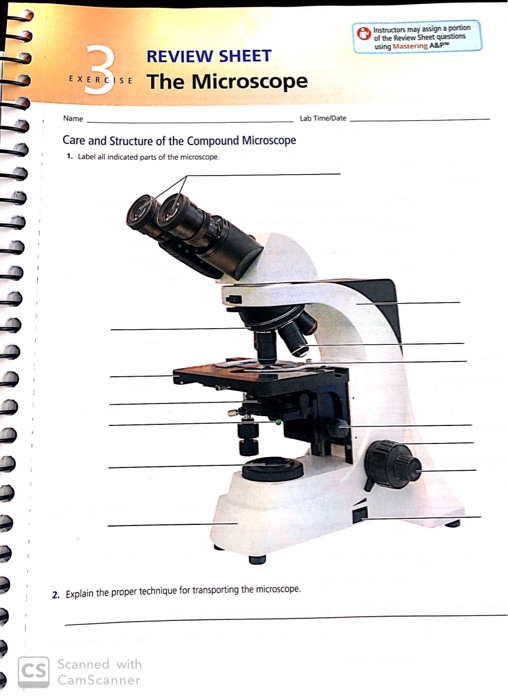
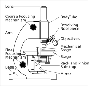
Parts of a microscope with functions and labeled diagram Head - This is also known as the body. It carries the optical parts in the upper part of the microscope. Base - It acts as microscopes support. It also carries microscopic illuminators. Arms - This is the part connecting the base and to the head and the eyepiece tube to the base of the microscope. Histology and Microscope Slide Labels Microscope Slide Labels. These specialty Microscope Slide Labels and matching End Labels are available in standard (thin) or pathology (tissue high) thickness, and square or round corner (RC). Permanent adhesive holds labels in place during use and long-term storage. Sheet Form Size is 5¼" x 8". Prices are per thousand labels. Slide Label
Microscope labels | Bill Todt Microscope labels. Unlabelled image: Lab Index: 101 Class Page: Identify the following parts of the microscope: Eyepieces Observation tube Nosepiece Objectives Stand Specimen holder Mechanical stage Stage controls Condenser Iris diaphragm Coarse focus Fine focus

Microscope images with labels
Microscope Slide Labels - LabTAG Laboratory Labels Chemical-resistant thermal-transfer labels designed specifically for the identification of glass microscope slides. When printed with our XAR-class solvent-resistant ribbons, these white or transparent histology labels, and the robust printout obtained, are capable of resisting xylene, toluene, alcohols, acetone, Methyl Ethyl Ketone (MEK), Dimethyl Sulfoxide (DMSO), as well as other solvents ... Amazing 27 Things Under The Microscope With Diagrams The tail is transparent and thus is difficult to detect under a low-power microscope. 23. Spirogyra under the microscope. Spirogyra is a green alga found mostly in freshwater in the form of green clumps. Spirogyra is unicellular, but because it clumps together, it can be seen in the pond even with our naked eyes. Explanation and Labelled Images - New York Microscope Company Another use of fluorescence imaging is Fluorescence Speckle Microscopy. It is a technology that uses fluorescence labeled macromolecular assemblies such as cytoskeletal protein to study movement and turnover rates. Fluorescence microscopy staining also is helpful in the field of mineralogical applications. It is routinely used for the study of ...
Microscope images with labels. Compound Microscope with labels Stock Vector | Adobe Stock Download Compound Microscope with labels Stock Vector and explore similar vectors at Adobe Stock. Adobe Stock. Photos Illustrations Vectors Videos Audio Templates Free Premium Editorial Fonts. ... Get 10 free Adobe Stock images. Start now. Get 10 free images. Unlock 200M+ assets in our full collection. Microscope picture label Flashcards | Quizlet Start studying Microscope picture label. Learn vocabulary, terms, and more with flashcards, games, and other study tools. Microscope Labeling - The Biology Corner The labeling worksheet could be used as a quiz or as part of direct instruction where students label the microscope as you go over what each part is used for. The google slides shown below have the same microscope image with the labels for students to copy. I often spend the first day walking students through the steps and having them look at a ... Parts of the Microscope with Labeling (also Free Printouts ... 5. Knobs (fine and coarse) By adjusting the knob, you can adjust the focus of the microscope. The majority of the microscope models today have the knobs mounted on the same part of the device. Image 5: The circled parts of the microscope are the fine and coarse adjustment knobs. Picture Source: bp.blogspot.com.
Microscope Illustrations and Stock Art. 67,723 Microscope ... Download Microscope images and photos. Over 67,723 Microscope pictures to choose from, with no signup needed. Download in under 30 seconds. Microscope Slide Labels - Histology Labels Our chemical-resistant microscope labels are compatible with a variety of thermal transfer printers including Cognitive CXT2-1300 and Zebra desktop printers. Adazon offers microscope slide labels in a variety of materials, sizes and shapes to meet the needs of your laboratory. Choose blank or pre-printed thermal transfer labels. Polarizing Microscope Image Gallery | Science Lab | Leica ... The position of the optical axis can be clearly determined with circular polarization. Right: Conoscopic image of the same calcite sample with linear polarized light. The calcite section is perpendicular to the optical axis. Images recorded with a DM4 P microscope using transmitted light, conoscopy, 63x N Plan objective, and polarizers. Microscope Images Labeled | Virtual Anatomy Lab VAL © 2023 by Yvonne Szymanski, Lisa Dubuc, & Bob Bucella Proudly created with Wix.com Wix.com
Simple Microscope - Diagram (Parts labelled), Principle ... The working principle of a simple microscope is that when a lens is held close to the eye, a virtual, magnified and erect image of a specimen is formed at the least possible distance from which a human eye can discern objects clearly. Magnification formula. The magnification power of a simple microscope is expressed as: M = 1 + D/F. Where Label the microscope - Science Learning Hub Use this interactive to identify and label the main parts of a microscope. Drag and drop the text labels onto the microscope diagram. eye piece lens: The lens you look through - normally 10x or 15x magnification. eye piece lens. coarse focus adjustment: Moves the lens up or down and adjusts focus. coarse focus adjustment. Microscope Parts and Functions With Labeled Diagram and ... Body tube (Head): The body tube connects the eyepiece to the objective lenses. Arm: The arm connects the body tube to the base of the microscope. Coarse adjustment: Brings the specimen into general focus. Fine adjustment: Fine tunes the focus and increases the detail of the specimen. Nosepiece: A rotating turret that houses the objective lenses ... Parts of Microscope, Function, Names & Labeled Diagram ... Condenser. The condenser is to focus the light, which passes from the microscopic illuminator to the specimen. This condenser is located just below the diaphragm. This diaphragm is one of the important parts of the compound microscope which will help to get an accurate and sharp image. The condenser has a magnification power of 400X and above.
Microscopic Images
Microscope, Microscope Parts, Labeled Diagram, and Functions Revolving Nosepiece or Turret: Turret is the part of the microscope that holds two or multiple objective lenses and helps to rotate objective lenses and also helps to easily change power. Objective Lenses: Three are 3 or 4 objective lenses on a microscope. The objective lenses almost always consist of 4x, 10x, 40x and 100x powers. The most common eyepiece lens is 10x and when it coupled with ...
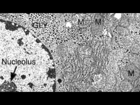
2.3.3 Identify structures from electron micrographs of liver cells - YouTube
PDF Label parts of the Microscope Label parts of the Microscope: . Created Date: 20150715115425Z

Science labs - Akers 6th Grade Team
Compound Microscope Parts - Labeled Diagram and their ... The term "compound" refers to the microscope having more than one lens. Basically, compound microscopes generate magnified images through an aligned pair of the objective lens and the ocular lens. In contrast, "simple microscopes" have only one convex lens and function more like glass magnifiers. [In this figure] Two "antique ...
35 Label The Compound Microscope - Labels Information List
Microscope Drawing And Label at PaintingValley.com ... All the best Microscope Drawing And Label 33+ collected on this page. Feel free to explore, study and enjoy paintings with PaintingValley.com. ... All rights to paintings and other images found on PaintingValley.com are owned by their respective owners (authors, artists), and the Administration of the website doesn't bear responsibility for ...

Gastrointestinal Tract - Liver Histology - Embryology
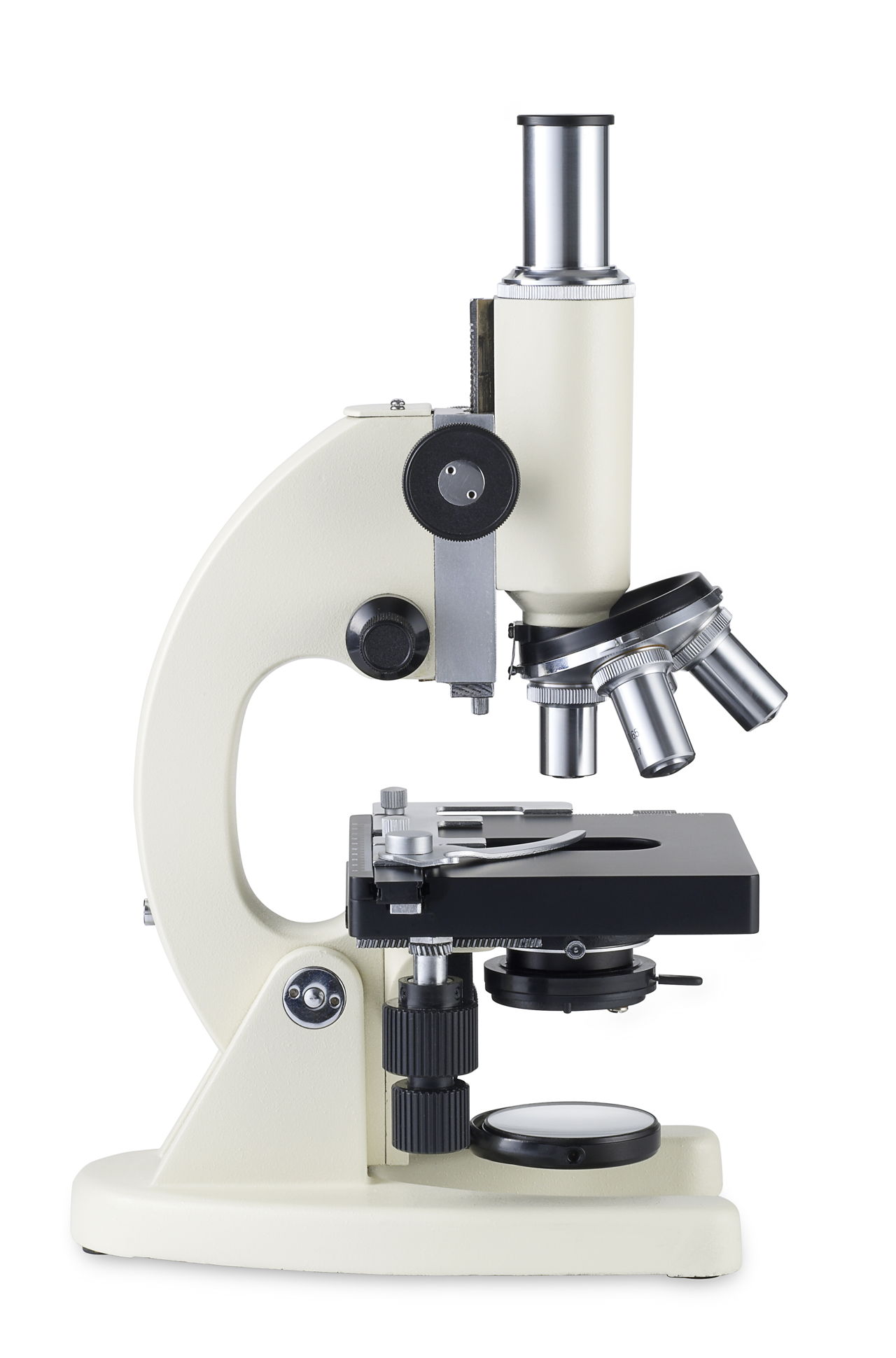
Microscope Types (with labeled diagrams) and Functions A compound microscope: Is used to view samples that are not visible to the naked eye. Uses two types of lenses - Objective and ocular lenses. Has a higher level of magnification - Typically up to 2000x. Is used in hospitals and forensic labs by scientists, biologists and researchers to study micro organisms. Compound microscope labeled diagram.
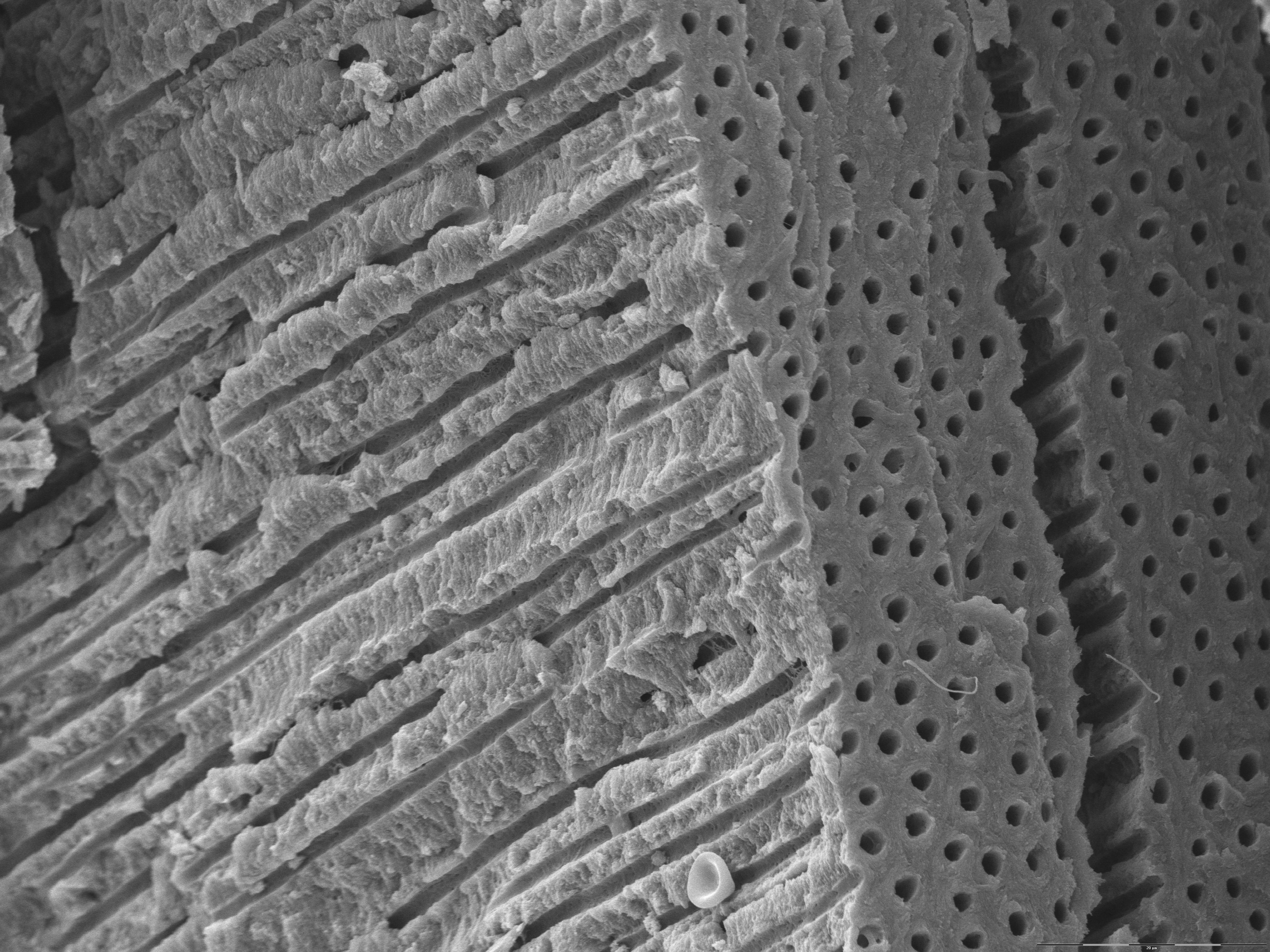
Dentine with dentinal tubules - Anatomicum.com
Microscope Drawing Easy with Label - YouTube In this video I go over a microscope drawing that is easy with label. There is a blank copy at the end of the video to review on your own. A great way to s...

Week 1: Microscope Usage & Snowflake Preservation - Inspiring a Love of Life-Long Learning
Compound Microscope: Definition, Diagram, Parts, Uses ... Compound microscope is a type of optical microscope that is used for obtaining a high-resolution image. There are more than two lenses in a compound microscope. Learn about the working principle, parts and uses of a compound microscope along with a labeled diagram here.

Microscope labeling quiz | 6th grade science | Pinterest
Microscope With Labels clip art | Microscope parts ... Jul 3, 2012 - Download Clker's Microscope With Labels clip art and related images now. Multiple sizes and related images are all free on Clker.com.
33 Label A Compound Light Microscope - Labels Database 2020
Microscope Slide Labels | Quality Materials | Order at PDC StainerShield® Slide Label Stain Resistant - Direct Thermal Synthetic Permanent 1" Core 7/8" x 7/8" White, 7200 per Roll, 1 Roll per Box. TDSS1-4-7878. $1,146.26. Add to Cart. Add to Compare. StainerShield® Slide Label Stain Resistant - Direct Thermal Synthetic Permanent 3" Core 7/8" x 7/8" White, 1800 per Roll, 4 Rolls per Box. TDSS3-1-7878.

Label the Microscope Part
Labeling the Parts of the Microscope | Microscope World ... Labeling the Parts of the Microscope. This activity has been designed for use in homes and schools. Each microscope layout (both blank and the version with answers) are available as PDF downloads. You can view a more in-depth review of each part of the microscope here.

Salisbury's Graduate Histology: Connective Tissue
Explanation and Labelled Images - New York Microscope Company Another use of fluorescence imaging is Fluorescence Speckle Microscopy. It is a technology that uses fluorescence labeled macromolecular assemblies such as cytoskeletal protein to study movement and turnover rates. Fluorescence microscopy staining also is helpful in the field of mineralogical applications. It is routinely used for the study of ...

Synapse Science Magazine: Weird and Wonderful: Hidden Horrors Returns!
Amazing 27 Things Under The Microscope With Diagrams The tail is transparent and thus is difficult to detect under a low-power microscope. 23. Spirogyra under the microscope. Spirogyra is a green alga found mostly in freshwater in the form of green clumps. Spirogyra is unicellular, but because it clumps together, it can be seen in the pond even with our naked eyes.
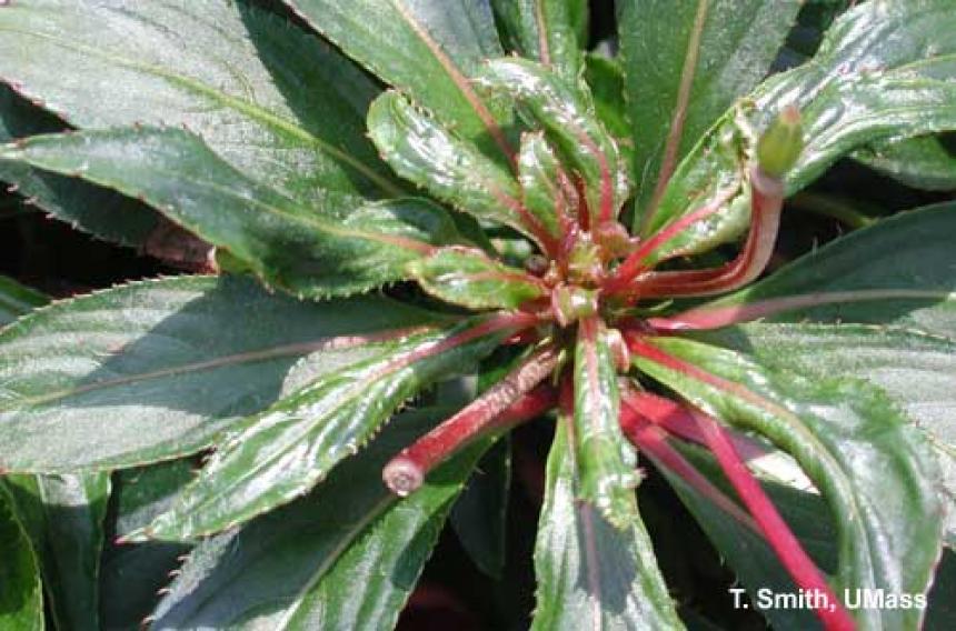
Broad mite feeding injury on New Guinea impatiens | UMass Center for Agriculture, Food and the ...
Microscope Slide Labels - LabTAG Laboratory Labels Chemical-resistant thermal-transfer labels designed specifically for the identification of glass microscope slides. When printed with our XAR-class solvent-resistant ribbons, these white or transparent histology labels, and the robust printout obtained, are capable of resisting xylene, toluene, alcohols, acetone, Methyl Ethyl Ketone (MEK), Dimethyl Sulfoxide (DMSO), as well as other solvents ...

35 Label The Microscope Game - Labels For Your Ideas

Biology Eleven: Looking at Agiospermae Reproductive organs. Also examine the leaf and stem cross ...
Microscope With Labels Clip Art at Clker.com - vector clip art online, royalty free & public domain
Label a microscope - Teaching resources

Microscope labeling
Post a Comment for "38 microscope images with labels"