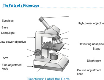
42 simple microscope diagram with labels
Kidney Cortex Histology Diagram Labeled - anatomyforme histology of the ... Kidney Cortex Histology Diagram Labeled - 17 images - kidney cortex, siu som histology crr, plate kidney cortex, kidney histology, › topics › medicine-andConfocal Microscopy - an overview | ScienceDirect Topics A standards document, which describes confocal microscopy and its influence quantities, has recently completed an ISO ballot as a final draft international standard (ISO FDIS 25178-607, 2018). A schematic diagram of a typical confocal microscope is shown in Fig. 15.1 (ASME B46-2009, 2010; Weller et al., 2012). Most examples of this method rely ...
Label-free chemical imaging of cytochrome P450 activity by Raman ... Raman microscopy has been used for label-free observation of biological molecules because it can directly detect molecular vibrations given by the intrinsic chemical structure of molecules 18.

Simple microscope diagram with labels
6 Stages of Plant life cycle in Detail with Diagram - Study Read The male part is called the stamen, which consists of a long filament called Anther, where the pollen matures. The pollen is disbursed onto the stigma of the female part, where the pollen gets attached to its base. As the pollen gets trapped, they transverse through the style to the ovary. The eggs in the ovary get fertilized with pollen. Plasma Membrane (Cell Membrane) - Genome.gov Definition. …. The plasma membrane, also called the cell membrane, is the membrane found in all cells that separates the interior of the cell from the outside environment. In bacterial and plant cells, a cell wall is attached to the plasma membrane on its outside surface. The plasma membrane consists of a lipid bilayer that is semipermeable. en.wikipedia.org › wiki › Electron_microscopeElectron microscope - Wikipedia An electron microscope is a microscope that uses a beam of accelerated electrons as a source of illumination. As the wavelength of an electron can be up to 100,000 times shorter than that of visible light photons , electron microscopes have a higher resolving power than light microscopes and can reveal the structure of smaller objects.
Simple microscope diagram with labels. Simple epithelium: Location, function, structure | Kenhub Simple squamous epithelium 1/4 Simple epitheliumhas only one cell layer where every cell is in direct contact with the underlying basement membrane. Generally, this type of epitheliumis found inside the body probably due to the fragile nature and forms the lining of the body cavities, blood and lymph vessels, heartand respiratory system. microscope | Types, Parts, History, Diagram, & Facts Optical microscopes can be simple, consisting of a single lens, or compound, consisting of several optical components in line. The hand magnifying glass can magnify about 3 to 20×. Single-lensed simple microscopes can magnify up to 300×—and are capable of revealing bacteria —while compound microscopes can magnify up to 2,000×. Brainstem: Definition, anatomy, parts, function | Kenhub The brainstem (brain stem) is the distal part of the brain that is made up of the midbrain, pons, and medulla oblongata. Each of the three components has its own unique structure and function. Together, they help to regulate breathing, heart rate, blood pressure, and several other important functions. Gram Staining: Principle, Procedure, Interpretation, Examples and Animation Procedure of Gram Staining. Take a clean, grease free slide. Prepare the smear of suspension on the clean slide with a loopful of sample. Crystal Violet was poured and kept for about 30 seconds to 1 minutes and rinse with water. Flood the gram's iodine for 1 minute and wash with water. Then ,wash with 95% alcohol or acetone for about 10-20 ...
Microscope Diagram - slide preparation biology4isc, anthrax microbewiki ... Microscope Diagram - 15 images - lycopodium, cell division of e coli with continuous media flow youtube, labelled microscope diagram gcse micropedia, give a well labelled diagram of compound microscope using of typical, Epithelial Cell Diagram Labeled - sensory summary smallcollation ... Epithelial Cell Diagram Labeled - 18 images - epithelial cells stock image g470 0052 science photo library, medical pictures info alveoli picture, epithelial cell function and structure, epithelia the histology guide, Light Microscope (Theory) - Amrita Vishwa Vidyapeetham Light microscope uses the properties of light to produce an enlarged image. It is the simplest type of microscope. Based on the simplicity of the microscope it may be categorized into: A) Simple microscope. B) Compound microscope. A) Simple microscope . It is uses only a single lens, e.g.: hand lens. Most of these are double convex or ... Basic Microscope Diagram - microscope diagram purposegames, images 01 ... Basic Microscope Diagram - 15 images - label the neuron clip art at vector clip art online, microscope diagram fill online printable fillable blank pdffiller, animal anatomy biology4isc, images 01 introduction and terminology basic human anatomy,
› books › NBK26880Looking at the Structure of Cells in the Microscope ... Many light-microscope techniques are available for observing cells. Cells that have been fixed and stained can be studied in a conventional light microscope, while antibodies coupled to fluorescent dyes can be used to locate specific molecules in cells in a fluorescence microscope. Living cells can be seen with phase-contrast, differential ... Microscope Diagram - cell division of e coli with continuous media flow ... Microscope Diagram - 15 images - give a well labelled diagram of compound microscope using of typical, bio tem biological transmission electron microscope university, labelled microscope diagram gcse micropedia, a compound microscope diagram micropedia, › articles › s41586/022/05028-xA physical wiring diagram for the human immune system | Nature Aug 03, 2022 · For imaging, a PerkinElmer Opera Phenix automated spinning-disk confocal microscope was used and each well of a 348-well plate was imaged at 20× magnification with 5 × 5 non-overlapping images ... how can a light microscope work - Be Refined Site Gallery Of Photos A simple light microscope manipulates how light enters the eye using a convex lens where both sides of the lens are curved outwards. Magnification can be achieved in a microscope by moving one or more lenses to different focal lengths. When looking through a microscope there are two ways that. Why can light microscopes produce color.
*Simple Squamous Epithelium*.........................(LOCATION: capillary walls, alveoli of ...
Coombs Test- Principle, Types, Procedure and Result Interpretation Procedure of Direct Coombs Test. Prepare a 5 % suspension in isotonic saline of the red blood cells to be tested. With clean pipette add one drop of the prepared cell suspension to a small tube. Wash three times with normal saline to remove all the traces of serum. Decant completely after the last washing.

All Saints Online: Diagram for Labelling: Microscope
Brigitte Zimmer Well Labelled Diagram Of Chlamydomonas. By Admin August 21, 2022 Post a Comment. Shipping a package with ups is easy, as you can print labels for boxes, paste them and even schedul…. Read more.

Download Labeled Diagram Of A Compound Microscope Gif - DirectScot
Chemical Laboratory Equipment Shapes and Usage | EdrawMax - Edrawsoft Step 3: To create a new lab apparatus drawing, go to [New] tab, find [Science and Education], and then click on [Laboratory Equipment]. This is where you'll find a wide range of templates to choose from and edit as needed. Step 4: To create a new one from zero, click on the "+" sign you can see in the picture above.

The Microscope: Create a Labelled Diagram | Teaching Resources
Epithelial Cell Diagram Labeled - epithelia the histology guide ... Epithelial Cell Diagram Labeled - 18 images - epithelial cells stock image g470 0052 science photo library, epithelial cells stock photos epithelial cells stock images alamy, types of epithelial cells stock image f031 6224 science photo library, apk 2100c chapter 4 tissues flashcards quizlet,

The Microscope: Create a Labelled Diagram | Teaching Resources
› en › microscopeFluorescence Resonance Energy Transfer (FRET) Microscopy Presented in Figure 3 is a Jablonski diagram illustrating the coupled transitions involved between the donor emission and acceptor absorbance in fluorescence resonance energy transfer. Absorption and emission transitions are represented by straight vertical arrows (green and red, respectively), while vibrational relaxation is indicated by wavy ...

White Blood Cells Diagram
Capillaries - Structure & Function Explained with Diagrams Structure of Capillaries. Capillaries have very thin walls comprised only of endothelial cells, which allows substances to move through the wall with ease. Capillaries are very small, measuring 5-10 micrometres in width. However, the cross-sectional area of capillaries within an average size muscle would be larger than that of the Aorta.

Minds Eye: August 2015
Testes: Anatomy, definition and diagram | Kenhub Put your knowledge to the test with our reproductive system labeling diagrams and quizzes. Each of the 200-300 lobules of the testis are filled with one to four highly convoluted seminiferous tubules which each course towards the mediastinum testis.

# 58 The immune system - Phagocytes | Biology Notes for A level
Differences between Light Microscope and Electron Microscope Light Microscope. Electron Microscope. Illuminating source is the Light. Illuminating source is the beam of electrons. Specimen preparation takes usually few minutes to hours. Specimen preparation takes usually takes few days. Live or Dead specimen may be seen. Only Dead or Dried specimens are seen. Condenser, Objective and eye piece lenses are ...

Microscope Diagram Labeled, Unlabeled and Blank | Parts of a Microscope | Science printables ...
filestore.aqa.org.uk › resources › scienceGCSE Science: Required practical activities - AQA Using a light microscope to observe, draw and label cells in an onion skin . Materials . In addition to access to general laboratory equipment, each student needs: • a small piece of onion • a knife or scalpel • a white tile • forceps • a microscope slide • a coverslip • a microscope • iodine solution in a dropping bottle.

anatomyforme: 2008-04-06
Different Size, Shape and Arrangement of Bacterial Cells When viewed under light microscope, most bacteria appear in variations of three major shapes: the rod (bacillus), the sphere (coccus) and the spiral type (vibrio). In fact, structure of bacteria has two aspects, arrangement and shape. So far as the arrangement is concerned, it may Paired (diplo), Grape-like clusters (staphylo) or Chains (strepto).

All Saints Online: Microscope Part Functions
Biology Schemes of work term 1-3 Form 4 - Educationnewshub.co.ke Draw and label a transverse section of a monocotyledonous stem. Examine transverse section of a monocotyledonous stem. Monocotyledo-nous stem, eg. tradescantia, microscope, Razors. KLB BK IV. PP111-2. 6: 3,4: Arrangement of tissues in a dicotyledonous stem. Draw and label a transverse section of a dicotyledonous stem.
Simple Microscope Labeled Diagram - Micropedia
The Compound Microscope Worksheet Answers - Division Worksheets Compound microscopes are more powerful than simple ones. It can be rotated to change the scale. Source: briefencounters.ca. ... Make a microscope on the parts and use the answer key on the worksheet and the parts on the blank microscope diagram. This worksheet asks students to name the different parts of a microscope and match key words to ...
All Saints Online: Diagram for Labelling: Microscope
Venn Diagram Templates | Editable Online or Download for Free Venn diagram template on different blogging platforms. Here is a 3 set Venn diagram that compares 3 popular blogging platforms; WordPress, Blogger and Tumblr. If you are starting a blog in the near future, this Venn diagram could be useful for you in making a choice between these platforms. Click on the image and use it as a template.
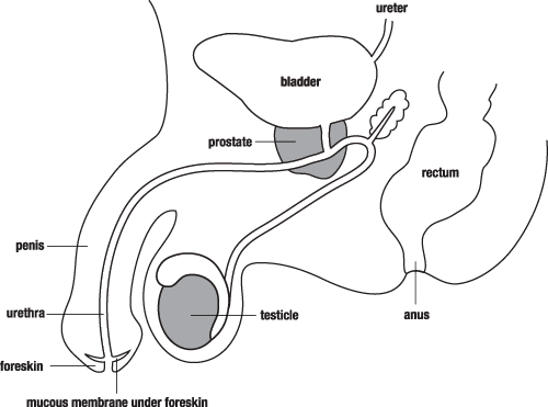
Male Reproductive System | Free Images at Clker.com - vector clip art online, royalty free ...
animal cell diagram labeled - Elouise Patel A diagram of a plant cell with the organelles labeled. Condensers are lenses that are used to collect and focus light from the illuminator into the specimen. They play a major role in ensuring clear sharp images are produced with a high magnification of 400X and above. Labeled diagram of a typical animal cell Nucleus.

31 Microscope Label And Functions - 1000+ Labels Ideas
animal cell diagram easy - Preeminence Log-Book Picture Library A simple diagram of an unspecialised animal cell labelled with numbers. Plant and Animal Cells by Angela Wagner at Christianbook. Most cells are very small. Popularly known as the Powerhouse. The control center of the cell. Ad Professional-grade PDF editing.

Microscope Clip Art at Clker.com - vector clip art online, royalty free & public domain
Spinal Cord Cross Section Explained (with Videos) Spinal Cord Cross Section. The spinal cord, which consists of the major nerve tract of vertebrates, runs down from the bottom of the brain through the passageway of the spinal column. This area is made up of all the nerve fibers that direct the reflex actions and convey the impulses that go back and forth to the brain.

A typical animal cell (as seen in an electron microscope) Medical Ima…
Hypodermis of the Skin Anatomy and Physiology - Verywell Health The hypodermis is the innermost layer of the skin. It stores fat and energy, pads and protects the body, attaches skin to the bones and muscle, and is very important in maintaining body temperature. This layer of the skin thins with age, increasing the risk for hypothermia or heat exhaustion. It provides shaping and contouring, and the ...
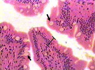
Simple columnar epithelium
en.wikipedia.org › wiki › FluorescenceFluorescence - Wikipedia Fluorescence is the emission of light by a substance that has absorbed light or other electromagnetic radiation.It is a form of luminescence.In most cases, the emitted light has a longer wavelength, and therefore a lower photon energy, than the absorbed radiation.
Post a Comment for "42 simple microscope diagram with labels"